


Coksartrosis- This is a hip joint arthrosis.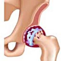 It develops gradually, several years prone to progress, it can be one-trailer and two-sided.It is signed by pain and restriction of movement in the wrist.In the later stages, the yellowish muscle atrophy is observed and limb shortening.The diagnosis is established on the basis of clinical symptoms and radiograph results.In the early stages of coxartrosis, conservative treatment.With the destruction of the compound, especially in patients youth and middle years, the operation (endoprosthetics) is indicated.
General information
Coxartrosis (osteoarthiness or deformation arthrosis blend of hips) is a degenerative-dystrophic disease.It usually develops at the age of 40 years and over.This can be the result of different injuries and joint diseases.Sometimes appears without obvious reason.Coxartrosis is characterized by a gradual progressive course.Conservative methods of treatment are used in early phases.In later stages, a common function can only be returned to operational.
In orthopedia and traumatology, cocsart is one of the most common arthritis.The high frequency of its development was created due to significant load on the hip joint and the widespread prevalence of congenital pathology - common dysplasia.Women suffer from coxartrosis a little more often than men.
COXARTS CAUSES
Primary (caused by unknown reasons) and secondary (developed as a result of other diseases) Arthreading hip joints.
Secondary coxartrosis can be the result of the following diseases:
- Dyplasia hip joint.
- The innate dislocation of the thigh.
- Pertes' diseases.
- Aseptic necrosis of thigh heads.
- Infectious lesions and inflammatory processes (for example, hip joint arthritis).
- Injuries (traumatic dislocations, hip fractures, pelvic breakers).
Coxartrosis can either be either one -sided or double -ed.With primary coxartrosis, an equal spine lesion (osteochondrose) and common knees (gonartrosis) are often noticed.
Risk factors
Among the factors that increase the likelihood of the development of coxarttrosis include:
- Constantly increased load on the compound.They are most often observed in athletes in people with excess body weight.
- Circulatory disorders, hormonal changes, metabolic disorders.
- Spine pathology (kyphosis, scoliosis) or stopping (flat feet).
- Older and senile age.
- Sedentary lifestyle.
Coxartrosis itself is not inherited.However, certain characteristics (metabolic disorders, skeletal structural characteristics and the weakness of the cartilage) child can inherit from their parents.Therefore, in the presence of blood cousins suffering from coxartrosis, the probability of the disease appears a little increased.
Patanatomy
The joints of the hips form two bones: Ileum and a boner.The head of the thigh is articulated by acetabulum Illian bone, forming distinctive "hinges".During the acetabulum movement, it remains motionless, and the boner moves in various directions, ensuring flexion, extension, abduction, adoption and rotational hips.
During the movement, joint surfaces of the bone unobstructed slack, thanks to the smooth, elastic and durable Croatian cartilage that cover the cavity of the rotary cavity and thigh head.In addition, Hijalin cartilage performs the function of shock-capation and is included in the redistribution of cargo during movement and walking.
In the shared cavity there is a small amount of shared liquid that plays the role of lubrication and provides food to Hijalin cartilage.The wrist is surrounded by a thick and strong capsule.Above the capsule are large female and gluteal muscles, which provide movements in the joint and, along with hyalin cartilage, are also shock absorbers that protect the wrist from injuries with unsuccessful movements.
With a coxartrosis, articulated fluid becomes thicker and more visual.The surface of the hyalin cartilage dries, loses smoothness, covered with cracks.Due to the roughness that appeared, cartilage during the movement is constantly injured on each other, which causes their thinning and deterioration of pathological changes in the wrist.As coxartost progresses, bones begin to deform, "customize" to increase pressure.The metabolism in the joint is worsening.In the later phases of coxartrosis, strong atrophy of the muscles of the painful limbs is observed.
Coxartrose symptoms
The main symptoms of the disease include pain in a common, inguinal end, thigh and common knee.Also, with kokesartrosis, stiffness movements and joint stiffness, home disorder, hromosy, hookish muscle atrophy and shortening of limbs on the lesionside.The characteristic coxartrosis characteristic is the restriction of the abduction (for example, the patient is difficult when trying to sit in a chair).The presence of certain signs and their seriousness depends on the coxartrosis phase.The first and the oldest symptom is pain.
InCoxartrosis 1. DegreePatients complain about periodic pain that occur after physical activity (running or extended walking).The pain is localized in the wrist, less often in thigh or knee.After a break, it usually disappears.Coxartrose 1. The degree is not broken, the movements are preserved in its entirety, there is no muscle atrophy.
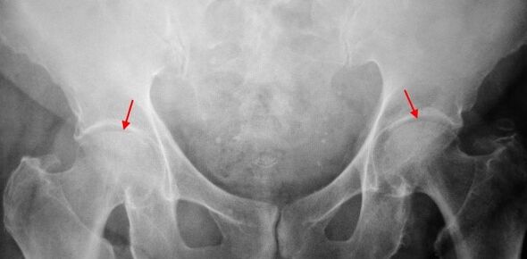
On X -Raj patient that suffers from coxartrosis 1. The degree is determined: moderately uneven narrution of a common gap, as well as the growth of bones around the outer or internal edge of acetabulum in the lack of changes from the head and the femur of the femur.
InCoxartrosis 2 degreesThe pain becomes more intense, often appear at rest, radiates into thigh and groin.After significant physical activity, the patient with coxartrosis begins to schedule.The scope of the joint movements decreases: abduction and inner thigh rotation is limited.
In X -Ray image for cooperation 2. degrees, significantly uneven narrowing of the common gap (more than half of the normal height).The federal head slung to the upward, deformed and increasing size, and its contours become uneven.The growth of bones with this degree of coxartrosis appear only on the inner, but also on the outer edge of acetabulum and go beyond the cartilage.
InCoxartrosis 3 degreesThe pain becomes constant, care for patients not only during the day, but also at night.The walk is heavy, when moving, a patient with coxartrosis is forced to use the reed.The volume of the movement in the joints is sharply restricted, the muscles of buttocks, hips and lower legs are atrophic.The weakness of the removal of the thigh muscles becomes the cause of the pelvic deviation in the front plane and shortening limbs on the painful side.In order to compensate for shortening, a patient suffering from coxartrosis, when walking, leaning the body in the sore throat.Therefore, in the center of the gravitational direction, the burden on the painful joint increases abruptly.
On the coxartrose radiograms 3. The degree revealed sharply of shared spaces, expressed the spread of the thigh head and multiple growth of bone bones.
Diagnostics
The diagnosis of coxartrosis is based on clinical signs and additional studies data, of which the main radiography is.In many cases, the X -Rays allows you to establish not only the degree of coextress, but also the cause of its phenomenon.Thus, for example, the increase in the corners of the neck-diaphone varnish, scenes and flattening of acetabulum indicate displasia and changes in the form of the Čutna bone indicated that coxartrosis is the consequence of Perres' disease or youthful epiphysiolysis.On the radiographs of patients with the coxarter, the changes can be disclosed to indicate injuries.
How other methods of instrumental diagnosis of coxartrosis, CT and MRI can be used.Computer tomography allows you to study the pathological changes in the matology structures, and the magnetic resonance provides the possibility of assessing soft tissue disorders.
Differential diagnosis
First of all, the coke-certainty should be distinguished from gonartrosis (osteoarthritis of a common knee) and osteochondrose of the spine.Atrophy of the muscle, which occurs in 2 and 3 phases of coxartrosis, can cause knee joint pain, which are often pronounced osaslija in the field of damage.Therefore, with the patient's complaints about the Knee Pain, a clinical (inspection, palpation of movements) is the study of the hip joint, and if coxarthrosis is suspected, to direct the patient to radiograph.
Pain for radicular syndrome (nerve root compression) for osteochondrose and some other spinal diseases can imitate pain with coxarter.Unlike Coxartrosis, when griping the root, the pain suddenly appears, after a failed movement, sharp turn, weight lifting, etc.It is localized in the area of the buttocks and spreads along the back of the thigh.The positive symptom of tension - strong pain when the patient tries to raise the correct limb, lying down on his back.At the same time, the patient takes his leg freely aside, while in patients with coxartrosis, abduction is limited.It should be borne in mind that osteochondrosis and coxartrosis can be respected at the same time, therefore, in all cases, a thorough patient examination is required.
In addition, CokeSartrosis is differentiated with trocharmis (boot loads) - aseptic inflammation in the area of glutel muscle attachment.Unlike coxartrosis, the disease is developing rapidly, within 1-2 weeks, usually after injury or significant physical activity.The intensity of pain is higher than in coxartrosis.Restrictions on movement and shortening of limbs have not been observed.
In some cases, with atypical during illness or reactive arthritis, coexart-resistant symptoms can be noticed.Unlike coxartrosis, with these diseases, the pinnacle of pain falls at night.Bol syndrome is very intense, can be reduced when walking.Morning rigidity is characteristic, which occurs immediately after waking up and gradually disappears within a few hours.
Coxartrose treatment
Treatment of pathology deals with traumatological orthopedics.The choice of treatment method depends on the symptoms and stages of the disease.Conservative therapy is performed at 1 and 2 phases of coxartrosis.During the coxartrosis deterioration period, injection blocks are used, not -steroidal anti-inflammatory drugs (pyroxy, indometacin, diclofenac, ibuprofen, etc.).It should be borne in mind that the drugs of this group has not been recommended for a long time, because they can have a negative effect on the internal organs and suppress the ability of Hyalin cartilage to return.
For the reconstruction of damaged cartilage for coxartrosis, funds from the hondroprotektor group (Hondroitin Sulfat, cartilage extract, etc.).To improve blood circulation and eliminate the spasm of small vessels, vasodilation drugs (zinarisin, nicotine, pentoxifilline, ksanhinol nicotine) are prescribed.According to the indications, muscle relaxants (drug muscles) are used.
With a stubborn pain syndrome, patients who suffer from coxartrosis can be prescribed by intra -ticular injections using hormonal drugs (hydrocortisone, triamcinolon, matrumor).Steroid treatment must be performed with caution.In addition, used by cocsartthrosis - heating fats that do not have a pronounced therapeutic effect, however, in some cases it relieves muscle cramp and reduce pain due to their "disruption".Also, with coxartrosis are physiotherapy procedures (radiant, ultrasonic therapy, laser treatment, UHF, inductermia, magnetotherapy), massage, manual therapy and therapeutic gymnastics.
Diet for coxartrose does not have an independent therapeutic effect and used only as a means of reducing weight.Reducing body weight allows you to reduce the load on the hip joints and, as a result, facilitate the coxartrosis course.In order to reduce the load on the joint, the doctor, depending on the degree of coxartrosis, can recommend walking with cane or crutches.
In later phases (with coke 3. degrees), the only effective method of treatment is operations - replacing the destroyed joint with endoprosthesis.Depending on the nature of the lesion, it can be used individually (replacement of the thigh heads) or two -poles (replacement and thighhead and rotary hollow).
The work of EndoProsthika for coxartons is done in the planned way, after the complete test, under general anesthesia.An antibiotic therapy is carried out in the postoperative period.The seam is removed for 10-12 days, after which the patient was prescribed for the ambulance.After endoprotetics, rehabilitation measures are necessary.
In 95% of cases, the surgical intervention for re-replacing the COCSArTtroZ joint provides a complete renovation of the limb function.Patients can work, actively move and even play sports.The average living life of the prosthesis, subject to all recommendations, is 15-20 years.Subsequently, another operation is needed to replace the worn endoprosthesis.